NIRSense Core Technology
All NIRSense systems include Near Infrared Spectroscopy (NIRS) and Photoplethysmography (PPG), two highly developed optical sensing techniques commonly used in medical devices. NIRS and PPG use deferent wave length light pulses that can pass through tissue. The characteristics of the reflected or scattered light can be analyzed to determine how much oxygen is in your blood, how much oxygen was delivered to critical organs and much more. Combining these and other sensing techniques into a light weight and durable device is what makes NIRSense truly unique.
 THE CARDIOPULMONARY SYSTEM
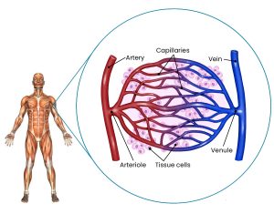
Muscles and organs require oxygen (O2) to function and stay healthy. O2 is delivered to the tissue by the blood through the complex vascular system. At the center of the system is the heart. The right side of the heart receives previously circulated blood and pumps it out to the lungs for reoxygenation. The left side of the heart receives fresh blood from the lungs and pumps it out to the tissue through arteries. After the oxygen is delivered to the tissue, the deoxygenated blood travels back to the heart through the veins, and the process starts over.
In the lungs, oxygen is attached to hemoglobin, a protein in red blood cells (RBCs). As fresh blood is pumped though the vascular system feed through smaller and smaller vessels eventually passing through the smallest known as capillaries. The hemoglobin deposits oxygen in the tissue where the cells use it as part of the energy generation process. The deoxygenated hemoglobin is carried away and back t the heart for recirculation. Near Infrared Spectroscopy (NIRS) can be used to detect and quantify the difference between oxygenated and deoxygenated hemoglobin producing the capability to measure oxygen delivery and consumption in specific tissue.
 TWO FUNDAMENTAL OPTICAL MEASUREMENTS
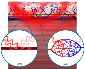
PPG measures optical changes in your arteries as they pulse oxygenated blood to your tissue. Comparing the optical changes in reflected light during and between pulses, and at different optical wavelengths allows for the calculation of pulse oximetry (SpO2). Analyzing the pulse timing yields pulse rate (PR) and, in some systems, respiratory rate (RR). Although PPG is typically measured on the fingertip or earlobe, it can be collected in any anatomical location where pulsatile blood can be measured.
NIRS measures optical absorption in both arteries and veins, and is frequently used for monitoring oxygenation in deeper tissues, particularly in organs like the brain and muscles. By comparing the reflected light from multiple wavelengths NIRS can detect changes in the concentration of oxygenated and deoxygenated hemoglobin and assess tissue oximetry (StO2). NIRS does not depend on the presence of a pulse, and can be measured on any vascularized tissue including muscle and bone giving more location flexibility. The depth of NIRS measurements is related to the distance between the light emitter and the detector. Typical NIRS systems can measure up to 2 cm deep, eliminating the effects of cold superficial tissue and making them more robust to movement and ambient light.
The fundamental difference between PPG and NIRS is that the former is a systemic measure of oxygen content in the arteries, while the latter is a specific measure of oxygen delivery and usage to the tissue of interest.
HOW IT WORKS
StO2 (Tissue Oxygen Saturation) measurement in a capillary bundle involves assessing the percentage of oxygen-saturated hemoglobin within a specific group of capillaries in a particular tissue or region. This measurement is typically performed using non-invasive near-infrared spectroscopy (NIRS) technology, which relies on the principles of light absorption and reflection to determine tissue oxygenation levels. A NIRS device consists of one or more light-emitting diodes (LEDs) that emit near-infrared light of specific wavelengths and one or more detectors that measure the light that is transmitted or reflected back from the tissue. The near-infrared light emitted by the LEDs penetrates the skin and underlying tissues and interacts with the hemoglobin molecules in the capillaries within the capillary bundle. Oxygenated hemoglobin (HbO2) and deoxygenated hemoglobin (Hb) have different absorption characteristics for near-infrared light. Oxygenated hemoglobin absorbs less light at specific wavelengths compared to deoxygenated hemoglobin. By measuring the amount of light absorbed by the tissue, the NIRS device can calculate the ratio of HbO2 to total hemoglobin (HbO2 + Hb), which is used to determine tissue oxygen saturation (StO2).
 NIRSENSE HAS COMBINED PPG AND NIRS INTO A SINGLE DURABLE DEVICE
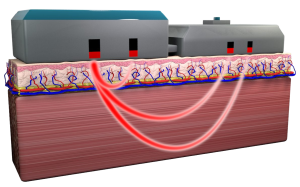
The depth of measurement in NIRSense systems is determined by the distance between the light emitter and the detectors. A shorter separation produces a shallower curve (channel) and measurement. Although most PPG devices measure the signal by passing light through the tissue to a detector on the other side (like a finger) NIRSense devices use the short channel to measure PPG from light reflected back to the detector. The deeper channels are used for NIRS and can reach as deep as 2 cm. The combination of these two technologies into one small, lightweight, durable, battery powered platform is a significant advancement in sensing technology.
PPG relies upon the pulsing component of arterial blood in order to record a measurement of oxygen delivery whereas NIRS relies on optical absorption and scattering from both underlying arteries and veins. These complementary sensing mechanisms give NIRSense the ability to determine a level of coherence between the systemic SpO2 and targeted StO2. And when the PPG signals fail due to motion, cold-induced vasoconstriction, skin pigmentation, or other confounding factors, NIRS can help maintain awareness about tissue perfusion and viability. The small form factor also provides great flexibility for placement of the device. In the event of a hemorrhage, the system may be placed on the deltoid or thigh. In traumatic brain injury (TBI) cases, the device could be placed on the forehead. Any area of the body where tissue oxygenation can provide patient health insights is accessible through the rugged form factor created to support US service members in the most challenging environments. This flexibility can provide additional patient comfort and improved equipment management in the Pediatric Intensive Care Unit (PICU), emergency transport, cath lab, or even surgery.
A COMPLEMENTARY PAIR
PPG and NIRS record complementary measures of cardiopulmonary function, and combining these two noninvasive optical measures can take advantage of each technique’s strengths to deliver a comprehensive monitoring of changes in cardiopulmonary function and local tissue oxygenation kinetics. For example, changes in oxygenated blood supply measured by PPG can be correlated with changes in tissue oxygenation demand and consumption measured by NIRS, providing a more comprehensive understanding of vascular responses in a variety of anatomical locations, across many use cases.
While NIRS has limited penetration depth and cannot distinguish between different types of tissues, PPG can provide information about superficial blood flow and pulsatile signals. By combining the two, you can gain insights into both tissue oxygenation (NIRS) and blood flow dynamics (PPG) at the same depth. This can enhance the understanding of physiological changes occurring in the measured area. The combination of PPG and NIRS allows for a more comprehensive assessment of tissue health. PPG provides information about cardiac parameters (e.g., heart rate and oxygen saturation), while NIRS offers insights into tissue oxygenation and hemodynamics. Together, they provide a broader perspective on a subject’s physiological status.
By monitoring blood flow and tissue oxygenation concurrently, this device can aid in the early detection of abnormalities or changes in a patient’s condition. For example, it can help identify circulatory issues, tissue hypoxia, or compromised perfusion at an earlier stage, allowing for timely intervention.